RadioDiagnosis
RadioDiagnosis
About the Department
Our founder director, Prof Ashok Kumar Mohapatra, inaugurated the Dept. with digital radiography, ultrasonography, and 64 Slice CT scan machine on December 27, 2013.
Since then, it has grown into one of the most advanced and well-equipped Dept. armed with state-of-the-art machines, handled by competent Radiologists and well-trained, experienced technologists. This Dept is ever ready (24x7) to serve the people of Odisha and people neighboring states. This Dept is a premier Dept of this institute catering to almost all modern imaging technique & playing a vital role in diagnostic & therapeutic aspects. To strengthen the academic activities in medical education, the Dept. had started postgraduate teaching programs in 2017. At present, 13 postgraduate students are undergoing training in all aspects of the fascinating field of radiology by a dedicated team of competent radiologists & technical staff. Currently, we have five senior residents working in our department.
We have also started a Post-doctoral certificate course (P.D.C.C.) in diagnostic neuroimaging in 2019 in the Dept.
The Dept. has also started BSc. M.T.R. (medical technology in radiography) course in 2016 with an annual intake of 13 students with its modern advanced equipment. The academics of the M.T.R. course at A.I.I.M.S., Bhubaneswar, are well designed to accomplish students in a highly competitive world.
We conduct the academic activities of the U.G., P.G., P.D.C.C., BSc M.T.R. regularly. The research activities & publication from this Dept are exceptional & deserves international repute.
Faculty
|
|
Facilities
DIGITAL SUBTRACTION ANGIOGRAPHY (Phillips Allura X.P.E.R. F.D. 20- biplane D.S.A.)
The D.S.A. system equipped with biplane detectors gives high-quality fluoroscopy and outstanding angiography and is one of the most advanced D.S.A. systems in the world. This dedicated neurovascular D.S.A. system with a large 40 x 40cms size flat panel tremendously decreases procedure time and radiation to the patient.
The superb image quality and advanced 3D tools help in confident decision-making during all kinds of endovascular procedures. High-resolution Imaging by the fixed X-ray system provides crisp, virtually distortion-free visualization of small details and objects to support endovascular interventions, including intracranial aneurysm coiling and carotid stenting procedures.
Features and applications
A dedicated Neurovascular Cath lab is available for all intracranial and extracranial
interventional procedures.
- Very high detector efficiency lowers the radiation dose for patients and staff.
- State of the art 3D-rotational angiogram (3D R.A.)
- Biplane configuration and 3D R.A. reduces procedure time significantly.
- C.T., like image acquisition (3D-CT), facilitates aggressive emergency management.
- Peripheral angiogram with bolus chasing significantly reduces examination time, radiation, and contrast doses.
- MAGNETIC RESONANCE IMAGING 3 TESLA (G.E. Discovery 750w)
- One of the leading M.R.I. systems with a wide bore and silent scan technology to benefit the patients for a comfortable scanning experience. High-definition structural and functional imaging capability in both neuro and cardiovascular applications provides excellent clinical diagnostic benefits.
- Features and applications
- Uses G.E.M. Technology (Geometry Embracing Method) to facilitate high resolution, high SNR whole-body imaging from the top of the head down to the feet.
- It is equipped with a powerful volume reconstruction engine (VRE) that enables real-time image generation, even when massive parallel-imaging datasets are involved.
- The Optix R.F. system enables high-bandwidth and high channel count reception with improved SNR than conventional M.R. receiver designs.
- Non-contrast 3D Arterial Spin Labelling perfusion imaging for various neuro and neurovascular diseases.
- High definition epilepsy and vessel wall imaging.
- Silent scan technology enables comfort scanning experience for the patients.
- Diffusion tensor imaging.
- Contrast-enhanced Time-Resolved Angiography for Neuro, Cardiac and Peripheral vascular diseases
- Capable of performing advanced structural & functional Imaging in both neuro and cardiovascular applications.
- MAGNETIC RESONANCE IMAGING (Siements 1.5T)
High-end 1.5T MR system, capable of performing one of the most comprehensive applications ranges available today. The unique combination of leading magnet and gradient technology and revolutionary image acquisition techniques provides unsurpassed clinical benefits. Excellent and efficient parallel imaging technique enhances the flexibility, accuracy, and speed of imaging examinations.
Features and applications
- The revolutionary parallel imaging (Tim - Total image matrix) technology.
- It is equipped with the most strong gradient in the industry - SQ-engine.
- Ultra-lightweight coils and low to the floor assures a high level of comfort for even very ill patients
- Advanced neuroimaging: Multinuclear M.R. Spectroscopy, diffusion tensor imaging, tractography, susceptibility-weighted Imaging, dynamic perfusion, functional M.R.I.
- Fast, comprehensive whole-body Imaging and central nervous system survey.
- 256 SLICE CT S.C.A.N.E.R. (Siemens Somatom Definition Flash)with dual source and dual energy tubes.
- The multislice C.T. has revolutionized cross-sectional Imaging with the exquisitely detailed multiplanar depiction of human anatomy and functional details. 256-slice dual-source with dual-energy tubes is one of the most advanced C.T. systems in the world. It delivers high image quality, dose efficiency, and rapid reconstruction times and can carry out a full-body scan in less than a minute.
Features and applications
- Expand clinical boundaries in neuro, cardiac, pulmonary, trauma, and pediatric Imaging
- Acquire 80mm of coverage in every rotation with sub-millimeter isotropic accuracy.
- Best-in-class reconstruction speeds using 3-D cone beam reconstruction.
- An efficient X-ray tube eliminates waiting times between scanning sequences and enhances workflow.
- Advanced whole-brain perfusion creates instant blood volume and blood flow maps to guide the timely treatment of ischaemic stroke.
- Comprehensive cardiac analysis with coronary C.T. angiogram and cardiac function analysis for complete care.
- Non-invasive cardiac perfusion assessment.
- Advanced 3D Imaging
- Carotid, cerebral, aortic and peripheral angiograms.
- Lung emphysema quantification for pre-surgical assessment.
- Lung nodule protocol to improve nodule detection sensitivity.
- Virtual endoscopy to perform and interpret exams more quickly.
- Bone mineral densitometry for quantitative analysis of osteoporosis.
- Body perfusion scan to assess the nature of the mass lesion.
- Dental CT and 3D face for enhancing surgery.
- 64 - SLICE CT Scanner (Siemens Somatom Definition AS)
- Dual slice (multislice) C.T. provides speed, resolution, image quality, reduced dose, and coverage optimal for an emergency C.T. study. Sub-millimeter slice thickness provides the high spatial resolution required for the visualization of small anatomy. Speed is sufficient to perform more aggressive studies like C.T. angiograms.
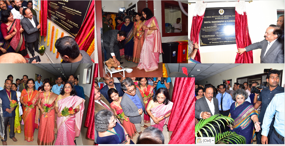
- Digital Radiography Fluoroscopy (Luminos DRF Max)
- To make reliable diagnoses, you need excellent visualization of all anatomical structures. Luminos DRF Max delivers clear images at the lowest possible dose. Plus, intelligent image acquisition and post-processing help keep examination and reading times short. The safety of patients and staff may be especially at risk during patient transfers to and from the system. Luminos DRF Max offers innovative design features and intuitive system operation to keep both patients and technologists safe.
- 3D Digital Mammography with Digital Breast Tomosynthesis (Hologoic, Selenia)
-
The Selenia Dimensions system performs the Imaging of tomorrow today. Streamlining workflow offers the following features.
- High-resolution display:
- Optimized face shield:
- FAST Paddle™ system:
- Streamlined tube head:
- Multiple procedure modes:
- 2D Mammography
- Genius™ 3D Mammography™ exam
- Low Dose Genius™ 3D Mammography™ exam
- 3D Mammography™ only
- 2D or 3D™ biopsy with the Affirm® breast biopsy guidance system
Ultrasound systems – (Philips, GE. Siemens)
- The premium performance ultrasound system captures crisp, high-resolution images even in technically challenging situations. It makes it easy to add 3D Imaging to any exam by removing the barriers to volume imaging, and a host of workflow enhancers facilitate faster exams, enabling perfect image optimization.
Features
- Obtain exceptional 2D and 3D image quality from a single transducer.
- Real-time, simultaneous Imaging is possible in two planes.
- Volume viewing on any PACS.
- Multi planar reconstruction of volume data is possible
- Enhancements in superficial soft tissue imaging.
Dedicated medical instrument navigation system guiding soft tissue biopsy and ablation
procedures.
- Appreciable reduction in examination time, pain, and fatigue related to the exam.
Digital and Computed Radiography (D.R. and C.R.) – (Allengers, Phillips)
- Flexible, high quality digital imaging solution enables seamless integration of the X-ray generator with the hospital or radiology information system. Innovative high end advanced flat panel detectors provide outstanding image quality, optimal workflow and operability.
Features
- Virtually unlimited positioning freedom.
- Auto-positioning.
- Software to enhance image quality and workflow.
- Appreciable reduction in examination time.
- Superb image quality and potential for dose reduction.
- Supports general radiology, including full leg, full spine, extremities, neonatal and pediatric applications.
- Optimizes cassette-based workflow with drop-and-go buffer.
- High throughput and fast preview of images.
- Small footprint - better management of available space.
- D.I.C.O.M. connectivity and integration.
Mobile Radiography (Skanray) – Three in number
Mini PACS (Picture archiving and communication system) – Siemens SYNGOPLAZA
- It makes the Department workflow more efficient, enables increased productivity, and serves hospital staff, patients, and referring physicians more effectively. It forms a comprehensive, fully integrated solution that meets all digital needs. The system features state of an art Picture Archiving and Communication System (PACS). Currently, we have 35 T.B. of storage capacity used to store D.I.C.O.M. images from all modalities of the Radiodiagnosis dept. Other departments can view the D.I.C.O.M. images of patients in their Department.
Services
- Interventional Radiological Procedures-
|
Vascular Interventional procedures- Therapeutic embolization procedures-
Renal artery pseudoaneurysm coiling
Sclerotherapy Diagnostic angiography
Hepatobiliary interventions –
Urological interventions
|
MRI-
|
CT Scan-
|
Ultrasonography and Doppler-
|
- Conventional radiology and fluoroscopy-
|
- Mammography-
|
- Dedicated services to COVID patients in admitted (ward and I.C.U.) patients and emergency services to COVID patients round the clock.
|
H.O.D.' S MESSAGE
It is indeed a proud privilege on my part to express a few words about the Dept
as the founder professor & head of the Dept. of Radiodiagnosis and Interventional
radiology.
The branch of medicine deals with applying medical imaging technology to arrive
at an accurate diagnosis to aid in patient management. It is the most rapidly
evolving branch of medicine. Radiologists are specialists who view and interpret
the images and identify the abnormality to diagnose a disease entity. In addition to
providing a diagnosis, radiologists also perform image-guided interventions, which
are minimally invasive therapeutic procedures to treat diseases.
Ever since the creation of the Department, this Dept has never looked back on
providing auxiliary services to the clinician for timely management of the patient.
Currently, this Dept is functioning with world-class faculty, paramedical staff and is
equipped with state-of-the-art machines to meet any challenge in the diagnostic and
emerging arena. With a very high patient load, a well-structured academic program,
and a lot of scope for research, the postgraduate students have an approachable and
highly professional departmental environment with a lot of content for excelling in
the educational and research fronts. The alumni emerging from the portals of this
Department always have a cutting edge above the rest wherever they go.
My long-term vision is to be recognized as the Dept in the international platform
while subserving the best service to the needy. The entire motto is to deliver timely,
efficient radiological diagnostic & therapeutic services to needy people apart from
working hand to hand with the clinician to facilitate the patient care delivery system
round the clock. The entire machinery is engaged in helping the clinician to arrive
at the diagnosis & therapeutic intervention.
Visionary augmentation of this Department is to provide better service to the people,
academic pursuit & research in all aspects to meet the scientific development
challenge.
-
Significant Achievements:
- The Department started performing therapeutic endovascular interventional procedures under Digital Subtraction Angiography (D.S.A.) guidance.
- We performed therapeutic endovascular coiling of hepatic, renal, splenic, gastroduodenal, left gastric, and superior mesenteric artery pseudoaneurysm in trauma and chronic pancreatitis patients.
- We performed transarterial chemoembolization (TACE) in hepatocellular carcinoma.
- We performed therapeutic glue/P.V.A. embolization of the splenic artery in cases of hypersplenism.
- Preoperative tumor embolization.
- Embolization of uterine artery pseudoaneurysm and A.V.M.
- Transjugular Liver biopsy (T.J.L.B.)
- Glue embolization of A.V.M.
The Department performed major therapeutic non-vascular interventional
procedures.
- We have performed more than 100 percutaneous transhepatic biliary drainages (P.T.B.D.) procedures.
- Biliary stenting procedures.
- Percutaneous drainage of a large urinoma in sick COVID positive patient under ultrasonography guidance.
- Percutaneous nephrostomy procedure for hydronephrosis in sick COVID positive carcinoma cervix patient under ultrasonography guidance.
- We performed pneumatic reduction of acute intussusception in a pediatric patient under digital fluoroscopy guidance.
- C.T. guided biopsies
- U.S.G. guided biopsies and F.N.A.C.s
Functional M.R.I. (fMRI).
The Department is successfully providing nonstop dedicated services in the form of bedside Ultrasound, bedside X-rays, CT and M.R.I. of admitted (ward and I.C.U.) patients, and emergency radiological services to COVID patients with the help of a dedicated team of radiographers and radiologists around the clock.
M.R.I. and C.T. Imaging of Mucormycosis cases in COVID patients
- Stereotactic breast biopsies
- Clip placement and wire localization in the breast lesions.
- Cardiac M.R.I.
| Sl No | Name | Designation | Department | Action | |
|---|---|---|---|---|---|
| 1 |
69.jpg) Dr. Nerbadyswari Deep (Bag) |
Prof. & Head | Radiodiagnosis | radiol_nerbadyswari@aiimsbhubaneswar.edu.in | |
| 2 |
 Dr. Suprava Naik |
Associate Professor | Radiodiagnosis | radiol_suprava@aiimsbhubaneswar.edu.in | |
| 3 |
 Dr Sudipta Mohakud |
Associate Professor | Radiodiagnosis | radiol_sudipta@aiimsbhubaneswar.edu.in | |
| 4 |
 Dr. Biswajit Sahoo |
Asst. Prof. | Radiodiagnosis | radiol_biswajit@aiimsbhubaneswar.edu.in | |
| 5 |
 Dr Tara Prasad Tripathy |
Asst. Prof. | Radiodiagnosis | radiol_tara@aiimsbhubaneswar.edu.in | |
| 6 |
 Dr Ranjan Kumar Patel |
Asst. Prof. | Radiodiagnosis | Radiol_ranjan@aiimsbhubaneswar.edu.in | |
| 7 |
 Dr. Manoj Kumar Nayak |
Asst. Prof. | Radiodiagnosis | radiol_manoj@aiimsbhubaneswar.edu.in |
ACADEMIC PROGRAMMES-
- M.D. Radiodiagnosis.
- M.B.B.S. teaching.
- BSc Radiography course- Medical Technology in Radiography (M.T.R.)
SPECIAL CLINICS/ LABORATORIES/INTEREST/COURSE-
Post-Doctoral Certificate course (P.D.C.C.)- in Diagnostic Neuroradiology
- RESEARCH AREAS:
|
Sl. No. |
Title of intramural Projects of the Department |
Funding Agency |
Name of PI |
From – To |
|
1 |
Role of multiparametric M.R.I. in differentiating benign and malignant causes of non-specific gall bladder wall thickening |
A.I.I.M.S. Bhubaneswar |
Dr. Sudipta Mohakud |
2018-2020 |
|
2 |
M.R. Imaging in C.N.S. Tuberculosis |
A.I.I.M.S. Bhubaneswar |
Dr. Suprava Naik |
2017-2019 (completed) |
|
3. |
Perfusion M.R.I. in intracranial space occupying lesions |
Non-funded |
Dr. Suprava Naik |
2018-2020 |
Thirteen (13) Thesis/ Dissertation of P.G. students going on.
P.G. Students:
- Current (13)
- Alumni: (1)
-
Senior Residents:
- Current (3)
- Alumni: (10)
-
M.T.R. Students
- Current (32)
- Alumini (20)
Major Activities
-
Clinical Activities-
- Routine and Advanced radiological services
- Ultrasonography and Colour Doppler Imaging
- Reporting of various modalities -X-Ray, CT, M.R.I.,
- Special procedures, Mammography
- Image-guided Interventional procedures- F.N.A.C.s,
- Biopsies, Percutaneous Aspiration and drainage, P.T.B.D. and stenting, PCN.
- Endovascular Interventions- Coiling, Glue embolization,
- Tumor embolization, TACE, etc.
- Emergency services
- Portable Radiography
- Academics-
- Interdepartmental academic meet
-
Intradepartmental academics-
- Seminar
- Film reading
- Journal club
- Case presentation
- Research-
- Intramural and Extramural projects
- Thesis work
- Paper publications
- Photo Gallery:


